Haematology Watch, Vol.4 , Issue 1.
SHORT COMMUNICATIONThe Forgotten Mechanism in beta-Thalassaemia major
Abdulsalam Abbas Hashim
Ineffective erythropoiesis in β-thalassemia major:
In general, red cell hemolysis or shortened survival can be largely compensated by effective bone marrow hyperplasia which in turn results in marked peripheral reticulocytosis that can parallel the level of anemia unless a co-morbid nutritional anemia or ineffective erythropoiesis exist which in turn limits the ability of bone marrow response.
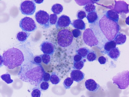
However, in β-thalassemia major, which is one form of hemolytic anemia, anemia results primarily from reduced and defective hemoglobin production. The presence of defective hemoglobin (excess of α-globin chains (1) that are highly unstable and precipitate in red cells precursors and progeny forming intracellular inclusions) in turn results in extravascular (producing splenomegaly) and intravascular (resulting in iron deposition in various body organs) hemolysis, and ineffective erythropoiesis (a mechanism that is crucial but usually much less appreciated). The level of reticulocytosis in this form of hemolytic anemia can vary in keeping with the degree of ineffective erythropoiesis.
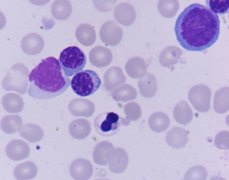
These images are from a 3 years old female with regular transfusion-dependant anemia since age of 3 months.
Hb 9.4 g/dl, WBC 5.0 * 109/l, Platelets 266 * 109/l
PB film revealed essentially a normchromic normocytic anemia with a smaller population of hypochromic microcytes. WBC and platelets are normal.
Hb electrophoresis was not performed before starting repeated blood transfusion due to the fact that this girl’s family is living in a poor rural area with no access to most of the diagnostic facilities. At presentation, the electrophoresis was unremarkable apart from mildly elevated HbF (4.1 %). However, this result should not be considered valid due to the fact of repeated blood transfusion.
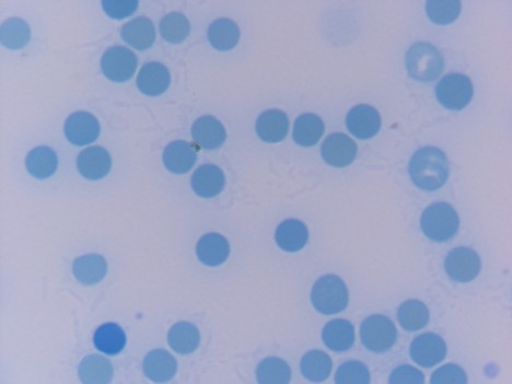
There was a marked reticulocytopenia, less than 0.1 % (Figure 1).